 1 / 9
Human Heart Coloring Pages: 30 Labeled Anatomy Studies of Chambers, Valves, Vessels and Blood Flow
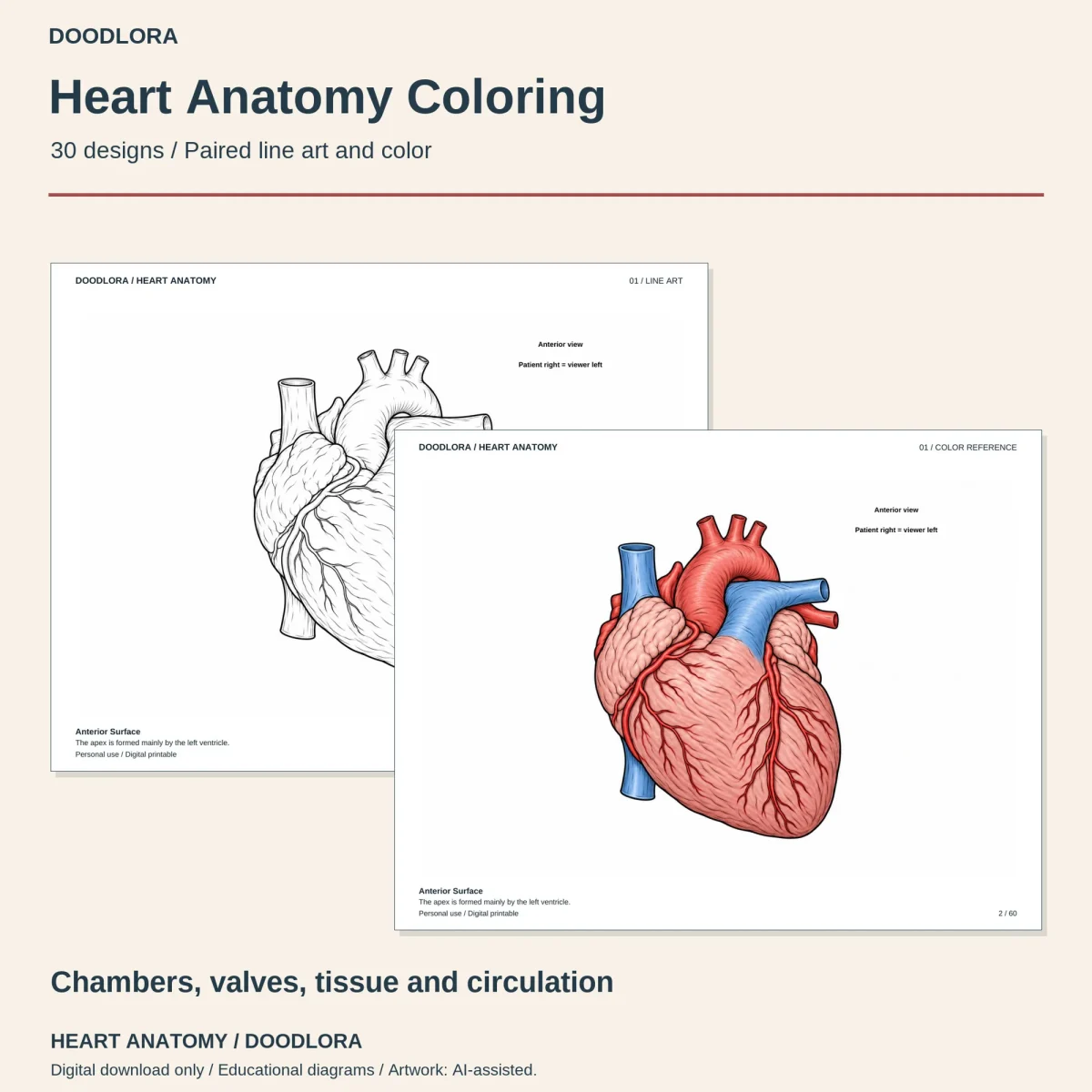
A 60-page printable book that pairs 30 heart anatomy studies with 30 color pages, line art first and its color version straight after. Most pages are labeled diagrams, with two realistic outside views at the start and four desk scenes with heart models at the end.
$3.99
Buy any 3 or more printables and save 20% at checkout.
- Download right after payment — link works for 30 days, up to 5 downloads
- Files arrive as a ZIP with your license PDF inside
- Secure checkout by Stripe · Refund policy
These human heart coloring pages come as one printable book: 30 designs, each drawn as black line art and then repeated in color on the very next page. Designs 03 to 25 have their parts labeled in print, such as Right atrium, Chordae tendineae or SA node, and every page carries a title and one sentence along the bottom edge.
What the 30 designs cover
| Designs | Topic |
|---|---|
| 01–02 | Realistic shaded views of the heart from the front and from the back |
| 03–08 | The four chambers, the inside of the right atrium, the right ventricle’s outflow, the left atrium with four pulmonary veins, the thicker left ventricle, and the wall between the ventricles |
| 09–13 | Valves: the three-leaflet tricuspid, the two-leaflet mitral, the aortic valve closed and open, the pulmonary valve, and the cords and small muscles that hold the leaflets |
| 14–16 | Coronary arteries and cardiac veins as shaded close-ups, then a diagram of tiny vessels running through heart muscle |
| 17–20 | The electrical conduction path, branching heart muscle cells, the three layers of the heart wall, and the sac around the heart |
| 21–25 | Blood flow: the lung loop, the body loop, a numbered 10-stop journey, filling versus emptying, and a cross-section of both ventricles |
| 26 | The heart between the lungs inside the chest |
| 27–30 | Desk scenes: cut-open heart models with books and pencils, a microscope with a magnified tissue circle, a model beside a stethoscope, and a table of whole and halved models |
Two drawing styles
Most of the labeled pages are simple outline diagrams with big open shapes to fill. The front and back views, the coronary close-ups, the muscle cells, the heart wall, the chest view and the four desk scenes are finely shaded drawings with far more line work, so expect those 11 designs to take longer to color.
Using the color pages
The color references keep to a small palette: blue for oxygen-poor blood, pink and red for oxygen-rich blood and muscle, yellow for fat on the surface. On designs 21 and 22 that blue and red is what shows the two loops, so it helps to keep the reference beside the line art for those. Print odd pages only for plain coloring sheets, landscape, one page per sheet at Actual Size. The A4 book matches the Letter one page for page.
Questions
- Why is some of the blood blue?
- The color pages use blue for oxygen-poor blood and red or pink for oxygen-rich blood so you can follow the two sides. It's a teaching color only; the start-here file states that blood is never literally blue.
- Which side is right on the diagrams?
- Right and left refer to the person, not to you looking at the page. On the front views the heart's right side sits on your left, and several pages print a small reminder; the back view on design 02 notes that there the sides line up with yours.
- Are the shapes and sizes accurate?
- They're simplified. Chambers, valves and vessels are drawn as clean diagram shapes, and some details are enlarged for clarity, such as the small vessels on design 16 and the sac around the heart on design 20.