 1 / 7
Digestive System Coloring Pages: 30 Labeled Diagrams from Mouth and Stomach to Intestines, Liver and Pancreas
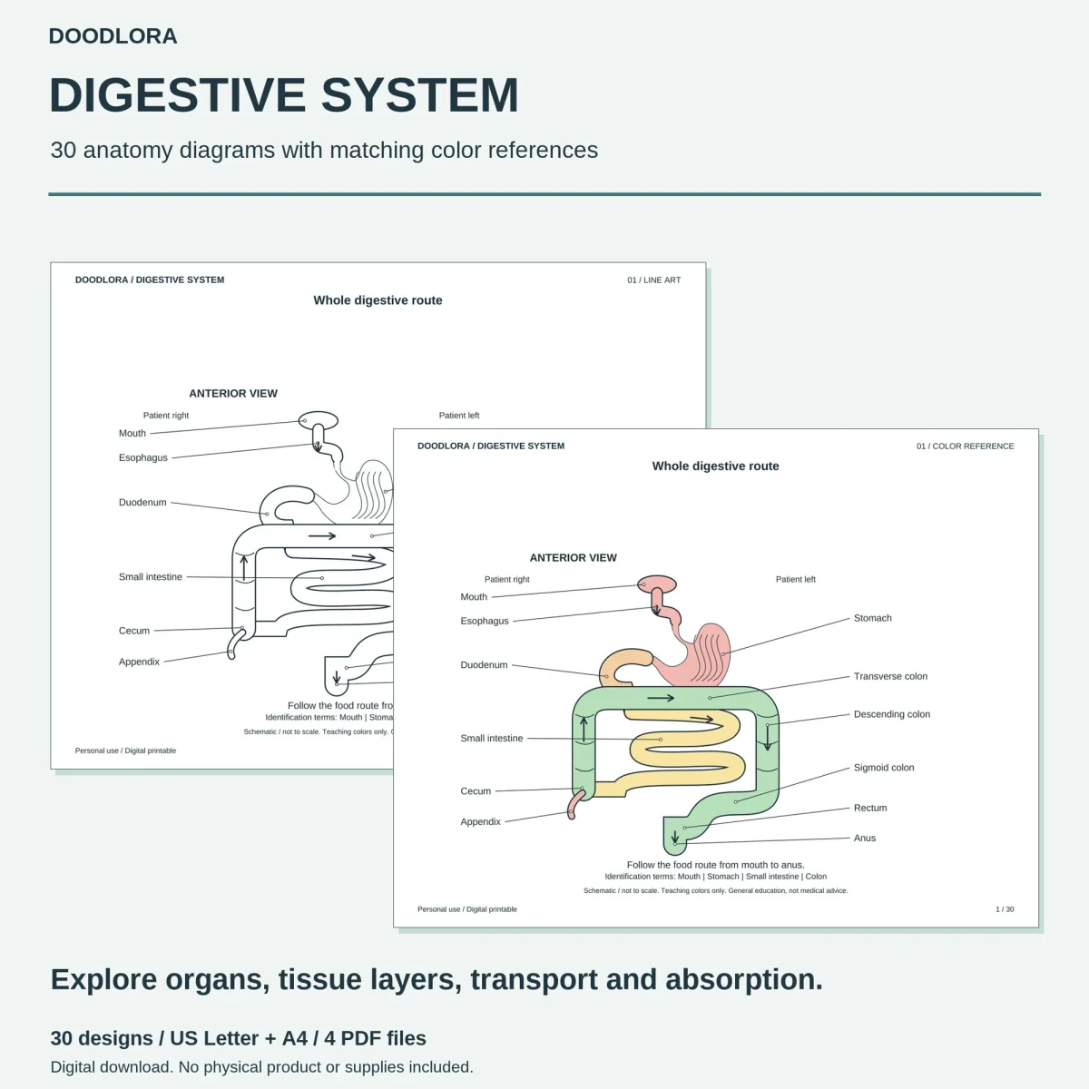
Thirty clean, labeled diagrams that follow food from the mouth to the colon, then cover the liver, gallbladder and pancreas. The line work is simple vector drawing with lots of white space, and a separate PDF shows every diagram in soft teaching colors.
$3.99
Buy any 3 or more printables and save 20% at checkout.
- Download right after payment — link works for 30 days, up to 5 downloads
- Files arrive as a ZIP with your license PDF inside
- Secure checkout by Stripe · Refund policy
These digestive system coloring pages are labeled diagrams, one per landscape page. Each part is named on the drawing with a thin leader line, and under the drawing are a one-sentence caption, such as “The stomach stores and mixes swallowed food,” and four study terms. The shapes are simple and outlined, and most pages leave plenty of white space around the diagram.
The 30 diagrams in order
| Pages | What they show |
|---|---|
| 01–03 | The whole route from mouth to anus, the layers of the intestinal wall, and the bile and pancreatic ducts meeting the duodenum, with an enlarged view of the opening |
| 04–08 | The mouth and esophagus: salivary glands, chewing food into a bolus, three phases of swallowing, the wave of muscle that pushes food along, and the esophagus in cross-section |
| 09–12 | The stomach: its regions, its three muscle layers, gastric pits and glands, and mixing and emptying through the pylorus |
| 13–19 | The small intestine: its three regions, folds, villi and microvilli, the inside of one villus, absorptive cells, how simple sugars and long-chain fats are taken up, and the mixing contractions |
| 20–25 | The large intestine: where the ileum joins the cecum, the route through the colon, its pouches and muscle bands, crypts and goblet cells, water absorption, and the rectum and anal sphincters |
| 26–30 | The liver and gallbladder, a liver lobule, pancreatic acini and ducts, bile breaking up fat, and the portal vein carrying blood from the intestine to the liver |
Step pages
Eight pages split the diagram into three side-by-side panels, and six of those are numbered steps, for example 1 chew, 2 mix, 3 form a bolus on page 05, or a large fat droplet turning into small droplets on page 29. Small arrows on these and a few other pages show which way food, blood or water moves.
Printing and the color file
The diagrams are vector line work, and the labels are real text you can select in a PDF reader. Print the coloring PDF on its own, in landscape at Actual Size, and keep the color reference open on a screen if you want a guide. Its palette sticks to soft pink, peach, yellow, green and pale blue, with the colon in green on the route pages and the airway in blue on the swallowing page.
Questions
- Are the four "Identification terms" at the bottom the answers?
- No. They're a short study list, not location labels. The parts are already named on the drawing itself, with leader lines pointing to each one, so nothing is left blank to fill in.
- Are the organs drawn to scale and in real colors?
- No. Every page notes that the diagram is schematic and not to scale, and that the colors are teaching colors. Organ sizes and cell shapes are simplified so each part is easy to see.