 1 / 10
Biology Coloring Pages: 30 Labeled Diagrams from Cells and DNA to Life Cycles and Food Webs
Thirty labeled diagrams that move from the inside of a cell to plants, fungi, small animals and the way energy and nutrients pass between them. The drawings are plain outlines with every label already printed, and each page has a numbered color version.
$3.99
Buy any 3 or more printables and save 20% at checkout.
- Download right after payment — link works for 30 days, up to 5 downloads
- Files arrive as a ZIP with your license PDF inside
- Secure checkout by Stripe · Refund policy
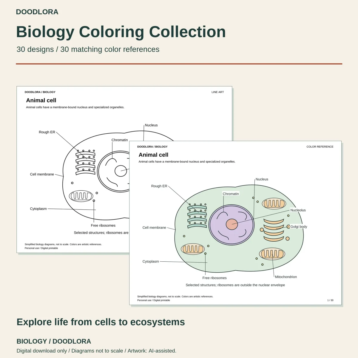
These biology coloring pages begin inside a single animal cell and end with fungi and bacteria breaking a dead leaf back down into nutrients for a new plant. Each of the 30 landscape pages is a diagram with its parts named in print and joined to the drawing by thin leader lines. The outlines are smooth and roomy, with simple ovals, rings and dots standing in for what you’d see under a microscope.
From inside a cell to a whole ecosystem
| Pages | What’s drawn |
|---|---|
| 1–7 | An animal cell, a plant cell and a bacterium, then close-ups of the nucleus, a mitochondrion with its folded inner membrane, a chloroplast and the cell membrane |
| 8–10 | Mitosis in four stages, particles spreading across a dashed divider, and DNA as a twisted ladder next to A–T and C–G pair boxes |
| 11–17 | A flower cut lengthwise, the cell layers inside a leaf, a pore with its two guard cells, a root tip, a stem cross-section, a bean seed and a seed sprouting in four stages |
| 18–20 | A fern frond with its spore clusters, moss with capsules on stalks, and a mushroom above its branching hyphae |
| 21–25 | The outside of a bluegill and a ground beetle, how a feather’s barbs branch, and the life cycles of the monarch butterfly and the green frog |
| 26–30 | A grassland food chain, a pond food web, plant roots trading sugars with fungi, cells building into a leaf, and decomposition |
Plants, fungi and small animals take up most of the second half. There are no human body pages in this set.
The words on every page
Each page carries a title, one sentence above the drawing and a caption below it. On page 10 the top line reads “In DNA, A pairs with T and C pairs with G,” and the caption explains that the ladder is a schematic. The vocabulary is real biology, so you’ll meet terms like cristae, thylakoid, palisade mesophyll and mycorrhiza. On the food chain and food web pages the arrows run from the food to the animal that eats it.
The labels mark a selection of parts, not everything in the structure. The animal cell gets nine of them, from the rough ER to the free ribosomes, and the start-here notes say plainly that the labels pick out selected structures only.
Busy pages and quick ones
Most diagrams leave wide open areas, like the big central vacuole on page 2 or the seed halves on page 16. A few take longer. The cell membrane on page 7 is two long rows of round heads with paired tails, the bluegill on page 21 is covered in a scale pattern, and the stomata page is a wall of six-sided cells.
The color reference keeps to soft greens, lavender, peach and pale blue. It colors the feather blue and the nucleus purple, so treat it as one way to tell the parts apart, not as what they look like in nature. Print the line-art file at Actual Size with the page turned landscape, and choose Fit to printable area only if an edge gets cut off. The A4 PDFs repeat the same 30 diagrams.
Questions
- Is there a blank version for labeling practice?
- No. The part names are printed on the line art and on the color reference alike, so the pages work as labeled diagrams to color, not as fill-in-the-label sheets.
- Are the sizes and colors true to life?
- No. Every page footer says the diagrams are simplified and not to scale, and the start-here notes call the colors artistic suggestions. The monarch life cycle page also notes that the four stages aren't drawn to their real body sizes.